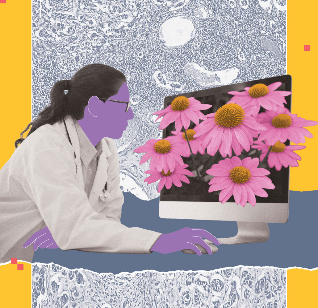
Biomedical engineer April Khademi is using AI in medical imaging to advance diagnosis, personalize treatment and potentially improve outcomes for patients with breast cancer and brain diseases like vascular dementia, Alzheimer’s disease and stroke. “As engineers, we try to build technology that helps society,” says Khademi, Canada Research Chair in AI for Medical Imaging, TMU professor of biomedical engineering, and principal investigator in the Image Analysis in Medicine Lab (IAMLAB).
“The AI tools our lab is developing for medical imaging help pathologists and radiologists to diagnose these conditions more accurately, precisely and quickly, allowing for better treatment decisions and quality of care,” says Khademi, who studied at TMU for her undergraduate and master’s degrees in electrical engineering.
AI-assisted pathology enhances breast cancer diagnosis
In Khademi’s recent study in Nature’s Scientific Reports, titled “AI improves accuracy, agreement and efficiency of pathologists for Ki67 assessments,” 90 international pathologists assessed Ki67 levels in breast cancer tissue with and without the aid of AI. The Ki67 biomarker is a crucial indicator for deciding treatment since a higher Ki67 proliferation score means tumor cells are dividing more rapidly, the cancer is aggressive, and the prognosis for survival is poorer.
“Ki67 scoring has traditionally been challenging for pathologists because counting thousands of tumour cells manually is laborious and subjective,” explains Khademi. “Pathologists may disagree on Ki67 scoring of the tissue, which can affect the quality and reliability of treatment decisions. Using AI helped pathologists be more accurate, they agreed more with colleagues, and it made them faster, which helps to streamline workflow and enhance patient care.”
When she was in her fourth year of the electrical engineering program at TMU, Khademi discovered in her capstone project that she could apply signal processing algorithms and machine learning to analyze digital medical images. “I found it super fascinating to build a new, cutting-edge technology that could potentially help patients,” she says.
AI imaging tools to personalize dementia diagnosis and tailor treatment
Since joining TMU as a professor in 2017, Khademi has collaborated with clinicians at several hospitals in designing AI algorithms. Her AI tools for medical brain imaging can detect patterns and extract meaningful quantitative biomarkers—such as the volume, size and shape of various brain structures—by analyzing very large amounts of neurological data from MRIs of patients with different types of dementia.
With vascular dementia and Alzheimer’s disease, for example, there is a clinical overlap that makes diagnosis challenging. In a 2023 study with clinical collaborators, Khademi’s patented AI tool identified biomarkers that could transform how clinicians diagnose the two distinct diseases. “Our AI tools can help to accurately diagnose patients earlier and offer them more personalized therapy and clinical drug trials.”
In another study with doctors at Sunnybrook and St. Michael’s hospitals, Khademi used a custom AI imaging tool to analyze MRIs from patients with mild cognitive impairment (MCI) and predict which patients would progress to Alzheimer’s to provide more tailored treatments. “Using these biomarkers, we predicted up to four years in advance whether a patient would convert from MCI to Alzheimer’s disease. This could allow clinicians to look at MCI patients and decide who to treat when and how with appropriate lifestyle modifications or medications tailored to the predictive diagnosis.”
These are still early days in terms of what Khademi can accomplish with AI-assisted medical imaging in transforming how doctors diagnose and personalize treatment for breast and possibly colon and pediatric cancers, and neurodegenerative diseases. “One of the best aspects of this research is to train bright minds and see what they do with the skills learned in my lab.”
Related stories


